
Macrophage Inhibition Factor
- Peritoneal exudates from antigen-sensitized guinea pigs, containing both
macrophages and lymphocytes are placed into capillary tubes. If the
sensitizing antigen is not added, the macrophages will migrate out of the
tube forming that fuzzy, mushroom-like head shown in the pictures.
Although the lymphocytes in the tube with the macrophages are committed
to one antigen (sensitized), they will not produce lymphokines until the
second or subsequent contact with the same antigen.
- The migration is inhibited when antigen to which the lymphocytes are
sensitized is added to the cell mixture. Macrophage Inhibition Factor
(Interferon) is released by sensitized lymphocytes in the capillary tubes,
responding to the presence of the sensitizing antigen. It is the MIF that
inhibits the macrophage migration not the antigen.
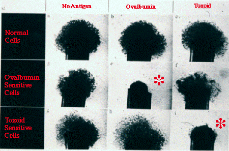
- Look at the photos. The top row shows the results when macrophages
and lymphocytes are collected from normal (unimmunized) guinea pigs. The
macrophages migrate freely with or without the antigens because the
lymphocytes are "seeing" the antigen for the first time; no MIF is
being produced. In addition, these controls show that any response by the
macrophages in the other tubes is independent of the nature of the
antigen.
- The inhibition of migration of macrophages from the tubes is only
seen in that tube which has both the ovalbumin sensitized
lymphocytes and the ovalbumin antigen, or the tube with both
toxoid sensitized lymphocytes and toxoid antigen.
 Return

Comments to MIPmaster: hthomp@lsumc.edu
Revised: August 1, 1996
URL:http://www.lsumc.edu/campus/micr/mif.htm
While every effort is made to ensure that this information is
up-to-date and accurate, the statements found on this page are for
informational purposes only.LSUMC WWW Publication Policy,
9/23/96