Case of the Month - April 2024
Yu Liu, MD; Liz Yang, MD; Ridin Balakrishnan, MD
A 52-year-old female with a history of abnormal uterine bleeding and an endometrial
mass detected on transvaginal ultrasound was admitted to the hospital for a hysteroscopy,
dilation and curettage, and myomectomy. Intraoperative findings of significance included
a 4.0 cm probable fibroid in the lower uterine segment. A resectoscope was used to
remove approximately 90% of this lesion. Multiple fragments of white-tan to pink-tan
fibroid-like tissue were sent to pathology for assessment. Microscopic sections demonstrated
a myometrial based neoplasm composed of spindled cells in vague fascicles surrounded
by delicate thin-walled vessels. However, areas of epithelioid morphology with clear
to eosinophilic and granular cytoplasm were also noted. Representative hematoxylin
and eosin (H&E)-stained sections at low and high power are illustrated in the Figure
1 below.
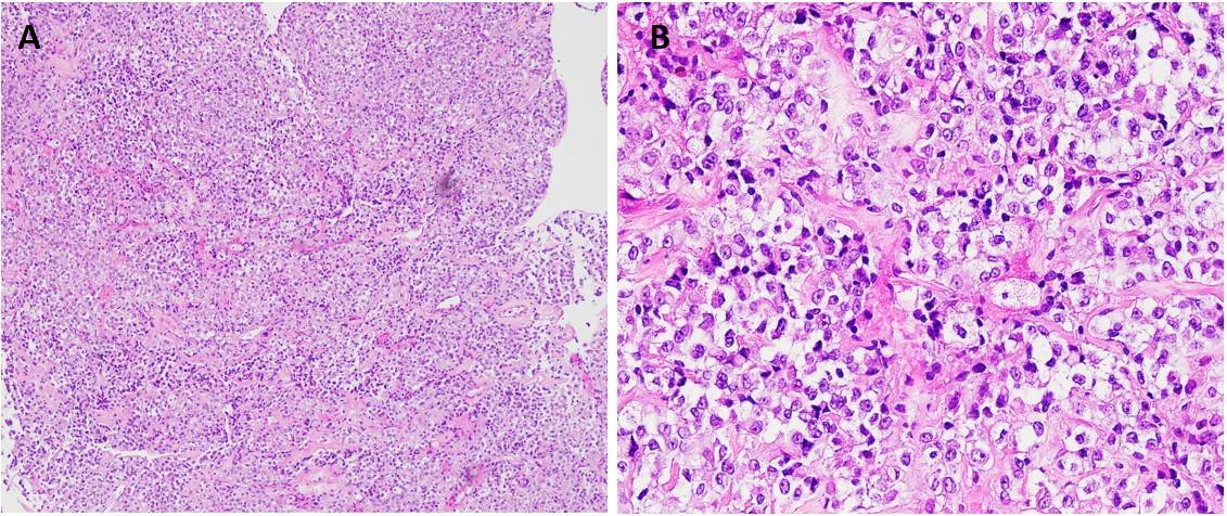
Figure 1: Epithelioid cells with clear to eosinophilic granular cytoplasm are arranged in sheets surrounded by delicate thin-walled vessels (A: H&E,100x; B: H&E, 400x)
By immunohistochemical (IHC) stains the lesional cells were patchy positive for Desmin (Figure 2A), ER/PR, p16, Calretinin, Melan A (Figure 2B) and HMB45 (Figure 2C), with rare expression of CK7. No significant expression of pan-cytokeratin, EMA, PAX8, p63, p40, GATA-3, S100, SOX10, CD117, and DOG1 was seen. P53 demonstrated wild-type staining. Some IHC stains are illustrated in the Figure 2 below.
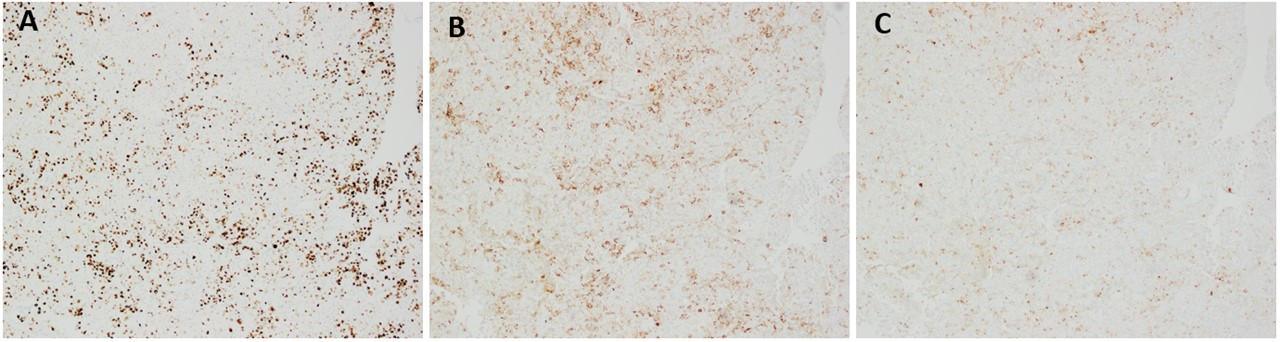
Figure 2: IHC stains (100x): (A) Desmin; (B) Melan A; (C): HMB45
Following the diagnosis, the patient returned to the hospital for a total laparoscopic hysterectomy and bilateral salpingo-oophorectomy. Gross examination of the resected specimen revealed a pink-tan, pedunculated, friable soft mass measuring 3.5 x 3.0 x 2.7 cm, attached to both the anterior and posterior left endometrium, occupying the entire endometrial canal. Representative H&E-stained sections from the mass are illustrated in Figure 3 (A, B: 200x) below. Most of the tumor in the resection resembled the spindled morphology of the lesion in the biopsy. Additionally, tumor cells focally demonstrated a perivascular radial distribution.

Figure 3: Tumor cells exhibit a perivascular distribution, with certain areas displaying spindled cells with eosinophilic cytoplasm organized into short fascicles (A, B: H&E, 200x)
The mass had an immunoprofile similar to the prior biopsy: SMA (patchy), Caldesmon (focal), Desmin (focal), HMB45 (diffuse), ER (focal), PR (patchy), and CD10 (focal). Diffuse expression of Cathepsin K (Figure 4A) was noted. Additional IHC performed for TFE3 (Figure 4B) was negative.
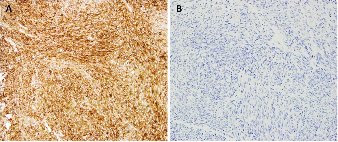
Figure 4: IHC stains (200x): (A) Cathepsin K; (B) TFE3.
What is the most likely diagnosis?
1. Epithelioid smooth muscle tumor
2. Alveolar soft part sarcoma
3. Perivascular epithelioid cell tumor
4. Malignant melanoma
5. Poorly Differentiated Endometrial Carcinoma
Click here for answer and discussion