
Medically important genera in the order Actinomycetales include Actinomyces, Mycobacterium, Nocardia and Streptomyces, all of which contain species pathogenic or potentially pathogenic for man. Some members of the Actinomycetales and the infections they produce have been grouped traditionally with the fungi and mycoses. Like the fungi, these "higher bacteria" form branching filaments, grow slowly, produce chronic infections, and elicit a cell mediated immune response in the host. However, Actinomycetales are true bacteria with procaryotic organization, and should not be confused with fungi.
ACTINOMYCES
Actinomyces israelii is the most common and important cause of human actinomycosis, a chronic suppurative and granulomatous disease characterized by abscess formation with peripheral spread to contiguous tissues and the formation of sinus tracts from the suppurative lesions. A. israelii, as well as the other agents of human actinomycosis, are indigenous to the oral cavity of man and may initiate infection following trauma to the oral tissues.
Other species of Actinomyces are found in the oral cavity, some of which may contribute along with other organisms to the pathogenesis of dental caries and periodontal disease (Actinomyces viscosus).
MYCOBACTERIUM
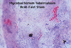
The most important member of the genus is Mycobacterium tuberculosis(hominis), the principal cause of human tuberculosis. Exposure to the tubercle bacillus is a significant occupational hazard to dental professionals. Tubercle bacilli are fairly resistant to drying, and survive for long periods in dried sputum. Since it is also one of the most resistant bacteria to disinfection, tubercle bacilli are the principal target of any rigorous procedure for ensuring sterility or protecting professionals from communicable disease. The emergence of strains of Mycobacteria that are resistant to one or more of the standard mycobacterial antibiotics makes exposure to this organism particularly worrisome.
Mycobacterial disease can also be caused by any of several non-tuberculous mycobacteria that are divided into one of four groups on the basis of growth rate, colony morphology and pigment production. Most strains of non-tuberculous mycobacteria are less pathogenic for humans than M. tuberculosis.
Mycobacteria are acid-fast bacilli, i.e., resistant to decolorization with an acidic solvent after primary staining with a basic dye. This property is due to the large amount of complex lipids in the cell wall. Identification of M. tuberculosis in clinical specimen is the only basis for a definitive clinical diagnosis of tuberculous infection. In active pulmonary tuberculosis, viable tubercle bacilli are shed into the bronchial secretions (sputum), which therefore offer the best potential source for recovery of the organism. If the concentration of M. tuberculosis is high enough, direct microscopic examination of stained sputum may reveal typical acid-fast bacilli. This would be sufficient for a presumptive diagnosis, to be confirmed by culture on an appropriate medium. (Note, other bacteria normally found in the oral cavity are not acid-fast.)
The mycobacteria also include the leprosy bacillus Mycobacterium leprae, or Hansen's bacillus, which has never successfully been cultivated on artificial lifeless media.
| Organism | Pigment | Temp. Range | Rate of Growth | Colony Type | Niacin Produced |
|---|---|---|---|---|---|
| M. tuberculosis | none | 37°C | weeks | rough | + |
| Group I Photochromogens M. kansasii | orange (light) | 24-37°C | weeks | smooth | - |
| Group II Scotochromogens M. scrofulaceum | yellow- orange (dark) | 24-37°C | weeks | smooth | - |
| Group III Non-chromogens M. avium | none | 24-42°C | weeks | smooth | - |
| Group IV Rapid Growers M. fortuitum M. phlei | if any, yellow- orange | 24-45°C | days | smooth or rough | - |
