
EXERCISE 14
ORAL ANAEROBES
I. INTRODUCTION
Indigenous non-sporulating pyogenic anaerobic bacteria are more widely involved
in the causation of infection than has hitherto been suspected. The indigenous flora
of man is heavily weighted in favor of the anaerobes by factors of 10:1 on the skin
and in the vagina, 100:1 in the oral cavity, and as much as 1000:1 in the large
intestine.
Life-threatening infections (e.g., lung or brain abscess, peritonitis, septicemia, septic
abortion, etc.) caused by endogenous pyogenic anaerobes are now frequently seen in
the clinical setting. The most accessible natural sources for these organisms is the
oral cavity where they constitute a major proportion of the normal oral flora, but
where they may also be directly related to the pathogenesis of destruction of the
teeth and supporting structures. Each of these conditions, of immediate concern to
the dental professional, may also be the source of metastatic systemic infection at
distant sites. It may seem inconsistent that strict anaerobes, highly sensitive to
oxygen could normally inhabit the oral cavity constantly exposed to air; however,
anaerobic conditions are maintained:
- by the presence of any necrotic tissue in periodontal and crevicular spaces, and,
- through the utilization of oxygen by commensal aerobic bacteria growing in
the same microenvironment.
Recovery of fastidious pyogenic anaerobes from clinical specimens requires correct
use of prereduced transport media for sample collection and, in the laboratory,
appropriate anaerobic environmental systems and special media for primary
isolation. Speciation of isolated strains is generally accomplished by gas-liquid
chromatographic analysis for fatty acids, which give elution patterns characteristic
for each strain of bacteria.
The experiment which follows will show the relative ease with which anaerobes
now can be cultured, the major groups of oral anaerobes, and the relative numbers
of those groups. Several different growth media will be used in this experiment.
They include a non-selective medium and 4 different selective media designed to
show different groups of oral anaerobic flora.
Special Anaerobic Media:
- Anaerobic Blood Agar Plate (ANABAP) (1 green stripe)
- ANABAP is a rich non-selective medium that will give the total cultivable
anaerobic microflora, including both facultative and strict anaerobes. It differs from
a standard BAP in that it is supplemented with hemin and vitamin K, two growth
factors commonly required by anaerobes, especially Bacteriodes species.
- Columbia CNA agar with 5% sheep blood (1 blue stripe)
- This medium contains colistin (C) and nalidixic acid (NA), two antibiotics active
against Gram-negative bacteria. Growth on this medium is a reflection of total
cultivable Gram-positive, anaerobic microflora.
- Columbia CNA agar with 5% sheep blood and metronidazole (2 blue stripes)
- Most strict anaerobes are sensitive to metronidazole, while most facultative
anaerobes are metronidazole-resistant. This medium therefore estimates the
Gram-positive, facultative microflora.
- Tryptic soy agar (TSA) with 5% sheep blood, hemin, vitamin K, and vancomycin
(1 red stripe)
- Tryptic soy agar is a standard growth medium sometimes used as a blood agar base.
By now you should recognize the functions of most of the additions to this
medium. Vancomycin is added because most Gram-positive bacteria are
vancomycin sensitive. Hence, this medium represents the total cultivable Gram-
negative, anaerobic microflora.
- TSA with sheep blood, hemin, vitamin K, vancomycin, and metronidazole
(2 red stripes)
- This medium estimates the Gram-negative, facultative microflora.
II. LAB WORK
PART I: PLAQUE SURVEY (Tues. 9/12)
Materials supplied: (work in groups of 4)
- 1 tube containing HUMAN PLAQUE (black cap)
- 3 tubes THIOGLYCOLLATE (white dot)
- 5 swabs, sterile
- 6 pipets, 1 ml
- 3 ANABAP (1 green stripe)
- 3 CNA + S (1 blue stripe)
- 3 CNA + SM (2 blue stripes)
- 3 TSA + SHKV (1 red stripe)
- 3 TSA + SHKVM (2 red stripes)
Procedure:
FIRST LAB PERIOD
- Label the three thioglycollate tubes 5, 50, and 500.
- One student in the group should transfer 1.0 ml of the plaque suspension
with a 1 ml pipet to the tube labeled 5. Mix well. Discard this pipet.
- Use a fresh pipet to transfer 1.0 ml from this tube to the tube 50. Mix well.
Discard this pipet.
- Finally, use another fresh pipet to transfer 1.0 ml from tube 50 to tube 500.
Mix well. Discard this pipet.
- After labeling all plates with name and either 5, 50 or 500, three of the
students in the group will pipet 0.1 ml of his/her dilution onto each of
his/her plates. One student will not have a dilution or plates. (SAVE
THESE DILUTION TUBES FOR EXERCISE 15.)
- Use one sterile swab for each dilution set. Spread the liquid over the entire
surface of each plate in that set (e.g., one swab for all plates labeled 5; one
swab for plates labeled 50, etc.).
- Place the plates where the instructor designates for anaerobic incubation at
37°C.
SECOND LAB PERIOD (Tues. 9/19)
- Scan all the plates to get an idea of the population distribution. Make a
quantitative estimate for each group by multiplying the number of colonies
on a plate by the dilution factor for that plate. The dilution factor for this
exercise is the dilution x 10 (because only 0.1 ml was plated). Select one plate
out of each dilution set to count. The plate must have more than 50 but
fewer than 200 colonies to give an accurate evaluation.
- Try to pick out colonies typical of individual organisms. Compare your
plates with the pure cultures of various anaerobes on display at the front
table. Look especially for the dark grey or black colonies of Bacteroides.
- Fill out the Report Sheet and hand it in TODAY.
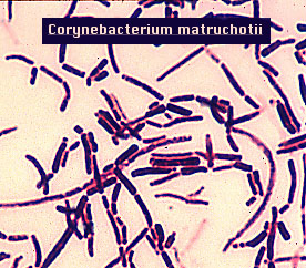
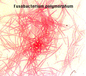
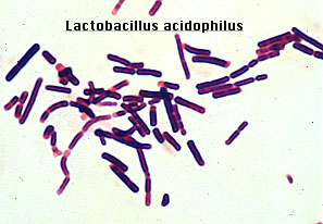
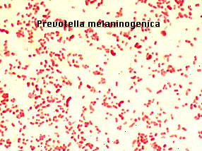
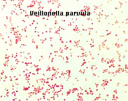
PART II: ORAL ANAEROBE DEMONSTRATION (Thurs., Tues., 9/14, 9/19)
- Observe the microscopic and macroscopic morphology of the oral organisms
on display at the front table.
- Fill in the following table with your observations. Accurate descriptions
will help with Part I of this exercise and later with the Lab Practical.

While every effort is made to ensure that this information is up-to-date and accurate, the statements found on this page are for informational purposes only.LSUMC WWW Publication Policy, 9/23/96