Of the numerous fungal genera known, approximately 40 contain species pathogenic for man. Most of these fungi are free-living saprobes, existing in the environment in decaying vegetation, bird and animal excreta, humus and soil. Infections in humans follow inhalation or traumatic implantation and are not usually spread from man to man. In the sense that infection is accidental, these fungi are opportunistic. However, while some of the "opportunists" can produce infection only in a compromised host, others are primary pathogens in normal individuals.
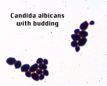
Fungi exist as either molds or yeasts; some types may be dimorphic and grow as both molds and yeasts. Molds are composed of tubular filaments called hyphae. These hyphae intertwine to form the mycelium, that is visible on natural substrates and on culture media at 25°C as the fungal colony. Yeasts are oval, unicellular fungi that reproduce by budding. Most are dimorphic. The parasitic form occurs in host tissues and on enriched media at 37°C and generally appear as yeast-like cells. The saprobic form occurs in nature and on special media at room temperature (25°C), and appears as hyphae. The parasitic form can be converted to the saprobic form by changing the environmental conditions.
The genus Candida comprises several dozen species of which at least eight have been reported to cause diseases in man. Candida albicans is the most important pathogen in the group. While Candida species are common members of the normal flora of the nasopharynx and gastrointestinal tract, they may cause severe cutaneous, mucocutaneous (e.g., oral thrush) and systemic infections. Conditions favoring infection are long-term antibiotic therapy, and suppression of cell-mediated immunity by chemotherapy or radiation. The organism is dimorphic; the yeast form occurs in the upper respiratory and intestinal tracts and in the vagina, while the hyphal or invasive form is found in mucosal and cutaneous lesions and in deeper tissues. Infection is initiated when a significant alteration in the mucosal or cutaneous tissue occurs, allowing the yeasts to produce hyphae and pseudohyphae that penetrates the epithelium.
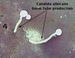
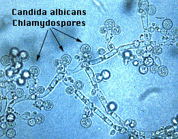
Isolation of yeast from oral swabs or blood specimen can be done on blood agar. Identification of C. albicans is based upon germ tube production, or production of terminal chlamydospores on hyphae and pseudohyphae grown on special media at 25°C, and on carbohydrate assimilation and fermentation patterns.
Histoplasmosis is caused by this dimorphic fungus. The fungus is found in nature or soil enriched by bird feces and in caves populated by bats. The organism is endemic in the Mississippi River Valley and other areas of the South.
Inhalation of spores produces a primary pulmonary infection. Depending on the dose of the inoculum and the overall condition of the patient, the infection by be self-limiting, involving only the lungs and less frequently, the spleen. Most often, lesions heal with calcification. The x-ray picture of histoplasmosis may be indistinguishable from that of tuberculosis. Presumptive diagnosis is based upon serologic findings, direct histopathologic examination of infected tissue and culture.
Blastomycosis, caused by this fungus is also endemic in this geographic area. Inhalation of spores usually induces a primary pulmonary infection similar to histoplasmosis. Chronic blastomycosis often spreads hematogenously from pulmonary sites to involve the skin.
Identification of the characteristic yeasts in sputum or skin biopsy samples establishes the presumptive diagnosis.
This dimorphic fungus is found in humus and rotting vegetation, on bark and thorns of shrubs, and in mulches such as sphagnum moss. Traumatic implantation of spores into the skin is the most frequent method of initiating infection. The most frequently observed type of sporotrichosis is the lymphocutaneous type, involving the extremities or other exposed areas of the body.
Diagnosis can be confirmed by culturing the organism at 25°C and observing thin septate hyphae with oval microconidia arranged in rosettes, and culturing at 37°C and demonstrating oval or football shaped yeasts.
Stock cultures on TSA plates:
A small amount of each yeast was suspended in individual tubes containing serum. The test tubes were incubated at 37°C for 2 to 3 hours. One drop of each yeast-serum suspension was placed on a glass slide along with India ink, to be observed as a wet preparation.
Examine each slide under the microscope for the presence of germ tubes.
| Organism | Germ Tube Formation |
|---|---|
| Candida albicans | + |
| Other Candida species | - |
Examine the stained microscope slide and photographs for the presence of large, round thick-walled chlamydospores.
| Organism | Chlamydospores Present |
|---|---|
| Candida albicans | + |
| Other Candida species | - |
