January 2024 Case of the month
Finding on Colonoscopy
Contributing Authors : Liz Yang, M.D. and Zhiyan Fu, M.D.
History
A 50-year-old person with no significant past medical history received a screening colonoscopy. A large pedunculated polyp, 1.5 x 1.4 x 0.9 cm, was found in the hepatic flexure and a complete polypectomy was performed during the procedure. The entire polyp was submitted for microscopic examination after serial sectioning. Representative sections stained with hematoxylin and eosin (H&E) and some immunohistochemical stains are demonstrated in the figures below.
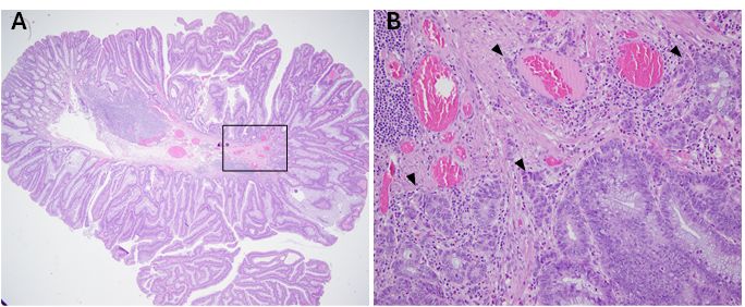
Figure 1 (A) Polypoid lesion is composed of conventional adenoma (H&E, 40x); (B) Small nests distributed at the polyp base (H&E, 200x)
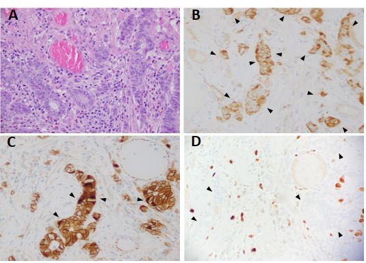
Figure 2 (A) Small nests distributed at the polyp base (H&E, 400x); (B, C, D)Immunohistochemical stains (400x) (B) Synaptophysin; (C): β -catenin; (D): Ki-67
Question
Based on the clinical information and the provided images, what is the most likely
diagnosis?
A. Colorectal invasive adenocarcinoma
B. Mixed neuroendocrine-non-neuroendocrine neoplasm (MiNEN)
C. Composite intestinal adenoma-microcarcinoid (CIAM)
D. Neuroendocrine tumor
E. Goblet cell adenocarcinoma (GCA)