
Faculty
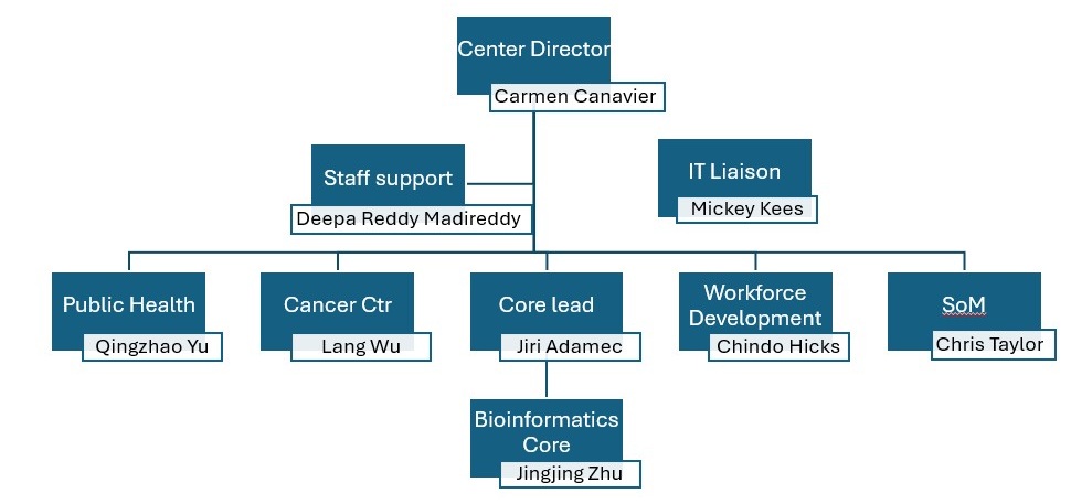

Carmen Canavier (Cell Biology)
We use self generated C, R and Python code to model the nonlinear dynamics of single neurons and networks of neurons. We are currently funded by NIH R01 DA041705 (Canavier PI) grant titled “A Dynamic Diversity of Dopamine Neurons” through May of 2029. We often use the simulation packages NEURON and NetPYne that are highly parallelizable. We run parameter sweeps at different parameter values and different initial conditions to check for robustness. We are currently one of the main users of the Tigerfish cluster. We also use genetic algorithms for to generate degenerate populations of models that fit a particular electrophysiological phenotype.
Our lab makes computational models of single neurons and of neural networks. We write the differential equations that describe the rate of change of membrane potential and other state variables then integrate them on Tigerfish. At the lowest level of modeling we model single ion channels using either a Hodgkin-Huxley formalism or a Markov model. Our models can be deterministic or stochastic. The next level of modeling is that of a single neuron. We sometimes use single compartment point neurons for simplicity. We also use multicompartmental models that model subcellular regions, such as the axon initial segment, the soma and different dendritic compartments separately. The next level of modeling is at the network level in which one or more populations of neurons are connected by chemical and sometimes by electrical synapses. We will take the modeling to the level of behavior by simulating classical conditioning and operant conditioning paradigms using Rescorla-Wagner reinforcement learning and Q learning for action selection. This work is in the context of drug addiction and builds on our current grant funding. The current funding is to build models that suggest the biophysical mechanisms underpinning the differences in how different populations of dopamine neurons, defined by their projection targets, integrate their synaptic inputs in distinct ways. The novelty of our behavioral modeling is that it will be the model of this (there are many) which models the firing of realistic dopamine neuron models and the release of dopamine in the different target regions. It will be a convergence of bottom up and top down models of dopaminergic signaling and its relationship to behavior. Current projects include modeling CaV2.3 in the context of improved therapies for Parkinson's disease.
The lab has a particular interest in how gamma oscillations (and oscillations in general) are generated in the brain. We model theta nested gamma in networks of hundreds of entorhinal cortical neurons, and many runs with different stochastic connectivity patterns and different initial conditions are required to characterize a single network. Parameter sweeps on these networks are very computationally intensive and benefit from parallelization.

Chris Taylor (MIP)
Dr. Taylor primarily uses open source, academically developed, free software for microbial community analysis (DADA2, phyloseq, Valencia, SpeciateIT, etc.), shotgun metagenomics analysis (biobakery, metaphlan, human, etc.) and machine learning.
Dr Taylor directs the Bioinformatics, Biostatistics, and Computational Biology Core of the “Louisiana Biomedical Research Network (LBRN)” P20 grant that supports and mentors a network of biomedical researchers throughout Louisiana. Dr. Taylor’s lab studies microbial communities and their interactions with the host. They are particularly interested in the interactions between reproductive tract microbiota and sexually transmitted infections. The goal of his R01 grant is to evaluate changes in the vaginal microbiome prior to occurrence of bacterial vaginosis and to study biofilm formation involving Gardnerella, Prevotella, and Atopobium. They use 16S rRNA gene sequencing and qPCR assays to assess changes in the vaginal microbiota leading up to bacterial vaginosis. His role as Co-Investigator and Subcontract PI involves supervising the qPCR assays and performing integrative analysis of the microbiome data. He uses machine learning methods to predict development of bacterial vaginosis from our microbial community sequencing data and qPCR measurements.

Chindo Hicks (Genetics)
Dr Hicks focuses on the development and application of bioinformatics and computational genomics methods and tools to analysis and integration of multi-platform multi-scale omics data generated using high-throughput genotyping, microarray, next generation sequencing and other related biotechnologies on cancer and other common human diseases and integration of these data with clinical information. He also develops and applies Machine Learning and Al algorithms to biomedical research and prediction of disease risks and threats. A third focus is data Mining and knowledge discovery. He uses Big Data analytics approaches to transform largescale omics data on common human diseases in diverse populations into liquidity and knowledge to improve human health and reduce health disparities.

Jingjing Zhu (Interdisciplinary Oncology)
Director of Bioinformatics core
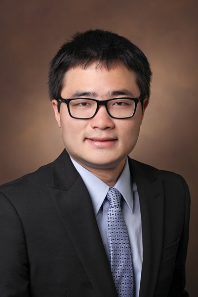
Lang Wu (Interdisciplinary Oncology)
Dr. Lang Wu’s research involves the epidemiologic investigation of genetic, molecular, nutritional, and lifestyle factors in etiology and prognosis of chronic diseases, especially cancer. His long-term research goal is to translate the gained knowledge for prevention, risk assessment, early detection, and prognosis prediction of human chronic diseases.
A main focus of Dr. Wu’s current research is to conduct integrative multi-omics (genomics, transcriptomics, methylomics, proteomics, metabolomics, etc.) studies to identify novel susceptibility genes and biomarkers for chronic diseases across populations. He has led several large transcriptome-wide association studies evaluating associations of genetically-predicted gene expression levels with risks of prostate, pancreatic, and breast cancers, which identified multiple novel susceptibility gene candidates for these cancers. He has also been investigating DNA methylation, protein, and metabolite biomarkers associated with human diseases using genetic instruments, which identified multiple novel biomarker candidates for prostate and pancreatic cancer. Furthermore, Dr. Wu is studying disease risk prediction using genetic, molecular, and other information.
Dr. Wu’s work involves machine learning (ML)/Artificial Intelligence (AI) for multi-omics analysis. The various omics data we analyze include but are not limited to below:
Genomics: Variant calling; SNP analysis; GWAS; Next-generation Sequencing including whole-exome and whole-genome sequencing etc;
Methylomics: DNA methylation and epigenetic regulation analyses;
Transcriptomics: RNA-seq; gene expression profiling;
Proteomics: Protein quantification and interaction networks;
Metabolomics: Metabolic profiling and pathway analysis;
We use multiple languages, software, and tools, including but are not limited to R, Python, Perl, and multiple Bioinformatics Tools (e.g., VCFtools, PLINK, BEDTools, SAMtools, BCFtools, ANNOVAR, METAL, EIGENSOFT, FOCUS, Seurat ,GCTA, and SMR and GsMap

Qingzhao Yu (Biostatistics & Data Science)
Dr. Yu directs the Research, Evaluation, Analysis and Design (READ). Population Health Center. They provide comprehensive services to support research and community health initiatives in Population Health. Services include but are not limited to: Study design, data management and visualization, statistical services, research methods, survey design and measurement development, participant recruitment in hidden and vulnerable populations, program evaluation and monitoring, environmental services, communication training, workshops, and seminars. Dr. Yu is funded by NIH R01 CA275089 “Interactions between ES-miRNAs and environmental risk factors responsible for TNBC progression and associated racial health disparities: a novel analysis with multilevel moderation influences”. She is an expert in machine learning. Research interests include Bayesian Modeling, Statistical Computation, Machine Learning, Spatial Analysis, Survey Data Analysis and Genetic Statistics.
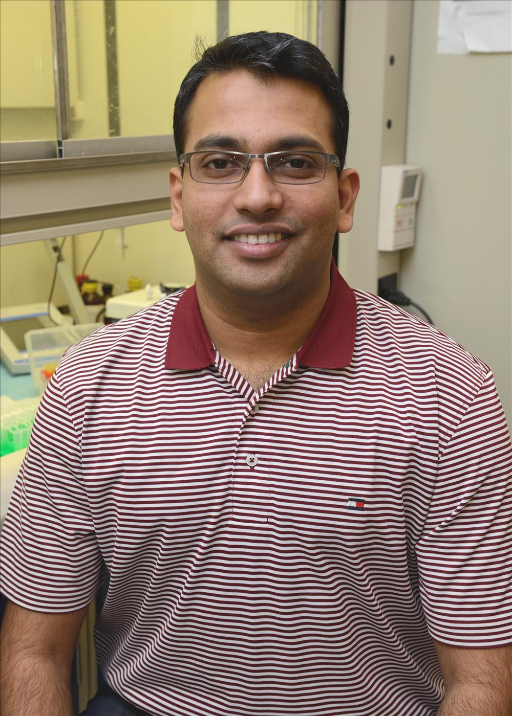
Bibo Bhattacharjee (Neuroscience Center)
Our lab utilizes a comprehensive set of tools described below.
I.10x Genomics Software Ecosystem for single-cell and spatial transcriptomics.
Cell Ranger converts raw single-cell gene expression (scRNA-seq) data into interpretable gene expression matrices, allowing us to identify the cell types and their transcriptomic states within our samples. Space Ranger This pipeline is for spatial gene expression data generated by 10x Genomics Visium platform is fundamental for understanding the spatial organization of gene expression changes within the brain, retina, heart regions, revealing how different transcriptomic and signaling pathways affect specific anatomical regions and cellular neighborhoods. Xenium Ranger: This is the processing pipeline for data from the 10x Genomics Xenium In Situ Analyzer, which allows us to pinpoint exactly where gene expression changes occur within individual brain neurons or glia, offering unprecedented detail on how drugs affect specific subcellular compartments or synaptic structures. Xenium Explorer: A dedicated visualization software for exploring the high-resolution spatial transcriptomics data generated by Xenium enables us to visually interrogate the precise spatial impact of our treatments on brain cellular architecture and gene expression, complementing our single-cell findings with crucial positional information.
II. R-based Bioinformatics Software
Seurat is used for single-cell RNA sequencing (scRNA-seq) data analysis including normalization, scaling, dimension reduction, clustering, differential expression, allowing us to identify cell types, discover shared gene modulation pathways, and analyze gene expression changes in response to treatments. Signac is used for chromatin accessibility analysis and enabling us to identify changes in chromatin accessibility, perform motif enrichment and link these epigenetic changes to gene expression. ArchR can integrate scRNA-seq data to explore gene regulatory networks, providing alternative or more advanced methods for dissecting the epigenomic landscape changes in our PAG samples. CellChat identifies significant ligand-receptor pairs between different cell populations allowing us to understand how different cell types in the PAG (e.g., neurons, glia) communicate with each other in response to pain. Nebula helps ensure that our identified differentially expressed genes are statistically robust, providing high confidence in our findings regarding gene modulation by pain and analgesic treatments. OmicScope allows multi-omic integration of our scRNA-seq and scATAC-seq data, helping us to connect epigenetic changes with gene expression modulation and ultimately the phenotypic response to pain and analgesics. Fiji (ImageJ) is an image processing package we use for analyzing microscopy images related to our spatial transcriptomics (Visium, Xenium) or for quantifying specific cellular features or protein expression from immunohistochemistry experiments that complement our omics data. DESeq2 provides a robust method for identifying genes significantly altered by our experimental conditions. Agilent Wave is used for RNA/DNA quality control, essential before performing sequencing.
III. MATLAB-based Bioinformatics Software
scGEAToolbox (Single-Cell Gene Expression Analysis Toolbox): A comprehensive MATLAB toolbox for single-cell gene expression data analysis that we use as an alternative or complementary platform to R/Seurat for our scRNA-seq analysis.
Other toolboxes we could potentially use: scInTime to model the temporal progression of cellular states in response to inflammatory pain and analgesic treatment within the PAG, even if cells are collected at a single time point. This could reveal how cells transition between different pain-sensing or pain-modulating states. scTenifoldKnk: single-cell network inference and perturbation analysis to explore the complex gene regulatory networks within any cluster of cells and understand how different drugs mechanistically influence these networks. scTenifoldNet: to use tensor decomposition for single-cell data integration and network analysis to compare and contrast the gene regulatory networks active in cells across different pain and treatment conditions.
Siyi Chen (Biostatistics and Data Science)
Dr. Chen’s research focuses on developing advanced statistical and computational frameworks for causal inference and integrative genomics in Alzheimer’s disease. Her work combines large-scale genomic, transcriptomic, proteomic, and imaging data to identify causal biomarkers and understand the molecular mechanisms driving neurodegeneration. Using approaches that integrate Mendelian randomization, Bayesian hierarchical modeling, nonlinear causal inference, and data-driven causal discovery, her research aims to map the complex pathways linking genetic variation, molecular traits, and disease outcomes. These methods provide a foundation for identifying biologically meaningful targets and uncovering nonlinear effects that traditional linear models often overlook.
Her analyses rely on intensive computation for data processing, simulation, and model estimation. The workflow involves R and C++ through RcppArmadillo, together with bioinformatics tools such as VCFtools, PLINK, and BEDTools for variant-level analysis, quality control, and data harmonization. High-performance computing resources are essential for handling large multi-omics datasets, running high-dimensional Bayesian inference, and exploring nonlinear causal models.

Michael Dunham (Otolaryngology)
Dr. Dunham collaborates with engineering and computer science faculty at LSU A&M on AI-assisted computer vision projects, including software development and medical instrument design. His group leverages LSU’s high-performance computing (HPC) cluster and the Advanced Machining and Manufacturing Facility (AMMF) for computational and prototyping needs. His lab is equipped with a multi-GPU workstation (Lambda Labs Vector) and medical-grade 3D printing capabilities (FormLabs).
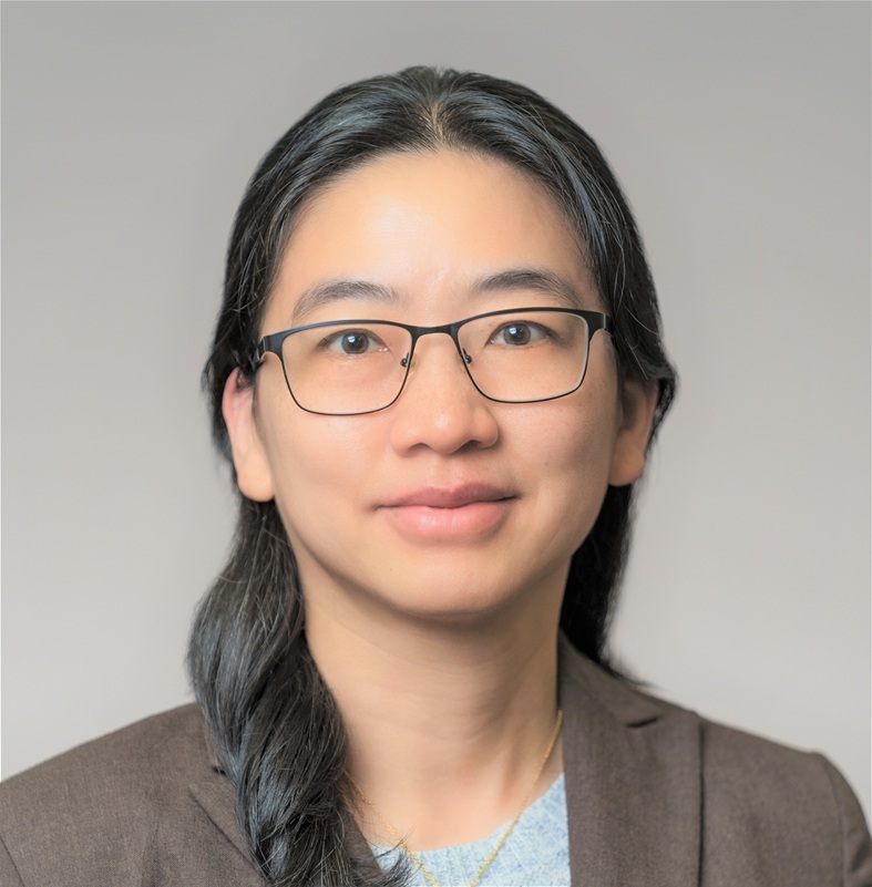
Huiyi Lin (Biostatistics & Data Science)
Dr. Huiyi Lin is a Professor in the Biostatistics and Data Science Program at Louisiana State University Health New Orleans. Her research focuses on the development of advanced statistical methodologies for the analysis of omics data, with particular emphasis on identifying gene-gene and gene-environment interactions that contribute to complex diseases. Dr. Lin has developed several innovative statistical methods and software tools for constructing polygenic risk scores and investigating SNP-SNP and SNP-environment interactions. Her current research involves large-scale genome-wide association studies (GWAS), particularly in prostate cancer, where understanding complex genetic interactions is critical for improving disease risk assessment and prognosis.
The analyses conducted in these studies involve massive genomic datasets and are highly computationally intensive. Efficient processing, storage, and modeling of such data require substantial computing resources, including high-capacity data storage systems and access to high-performance computing (HPC) clusters. These resources are essential for conducting large-scale genetic analyses and advancing discoveries in precision medicine and cancer research.
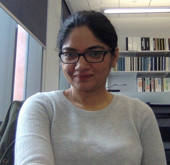
Rajani Maiya (Physiology)
Dr. Maiya’s lab leverages high-resolution transcriptomic technologies—including single-nucleus RNA sequencing (snRNA-seq) and spatial transcriptomics—to uncover cell type–specific molecular adaptations underlying substance use disorders and chronic pain. These approaches generate large and complex datasets that require powerful computational infrastructure for analysis and interpretation.
To this end, the lab relies extensively and almost exclusively on the LSUHSC high-performance computing servers for all stages of our snRNA-seq data analysis pipeline. This includes processing raw sequencing files, conducting quality control, generating count matrices that quantify gene expression per cell, and performing downstream analyses. The lab uses the R package Seurat for normalization, dimensionality reduction, clustering, and differential gene expression analysis. In addition, they utilize a suite of tools for data visualization and interpretation: DESeq2, Limma, EdgeR, , CellRanger, SpaceRanger, ScanPy, and ggplot.

Steffy Manjila (Neuroscience Center)
Dr.Manjilla’s laboratory employs tissue clearing and light sheet microscopy to visualize cell types in whole organs, particularly mouse brains, and spatial transcriptomics using the MERFISH-based MERSCOPE platform. These approaches generate exceptionally large datasets that exceed the capacity of standard workstations and require high-performance computing (HPC) for efficient processing.
Each whole-brain imaging experiment produces 100 GB to 1 TB of data at 0.4 µm XY and 2 µm Z resolution. These datasets are then analyzed and registered to a reference brain atlas using deep learning or machine learning–based pipelines with custom-codes in MATLAB and Python.
We are now advancing to submicron-resolution whole-brain imaging (0.5 µm XYZ) to reconstruct microglial and vascular architecture, which are highly relevant to aging, neuroinflammation, and disorders such as hypertension and stroke. At this scale, each brain generates ~4 TB of raw data per imaging channel, which cannot be processed feasibly without HPC infrastructure.
In parallel, the MERSCOPE spatial transcriptomics platform generates ~2 TB per experiment, with downstream analyses requiring specialized tools such as Scanpy and Squidpy specialized for spatial omics analysis. These tasks demand large memory and parallel processing, which only HPC can provide.
Amirsalar Mansouri (Cancer Center)
As a member of the Adamec Lab and the Proteomics and Metabolomics Core (PMC) core, Dr. Mansouri provides processing and analyzing high-dimensional mass spectrometry–based datasets. The core converts raw omics data into clean and interpretable results that are delivered to colleagues for downstream biological interpretation. The core also provides help with further statistical and functional analysis. The following are examples of open-source software and tools utilized:
Proteomics: MaxQuant, Perseus, FragPipe, MSFragger, Skyline
Metabolomics: MZmine, MS-DIAL.
Lipidomics: MS-DIAL, MetaboAnalyst, LipidLynxX.

Michael Maristany (Radiology)
Dr. Maristany recently became chair of radiology and wants to utilize machine learning to address problems in radiology. One project is to automate the determination of which patients are in shock and which are not. The data are CT images of patient’s abdomen and pelvis. He envisions a two-step process. First, the images need to be segmented to extract relevant features such as aorta diameter. Then the data needs to be separated into two groups, hopefully corresponding to shock versus no shock, possibly using UMAP.

Jason Middleton (Cell Biology)
Dr. Middleton is engaged in a collaborative project titled “Correlation Between Local Field Potential (LFP) Rhythms” with Lora Kahn and Julia Staisch (Ochsner), using neural recordings from Parkinson’s patients undergoing deep brain stimulation (DBS) electrode implantation. By extracting bandpass-filtered LFPs and analyzing dynamic power fluctuations, the project aims to uncover how cross-band correlations—such as between theta (4–8 Hz) and gamma (30–70 Hz) rhythms—relate to symptom expression. This work has potential to inform personalized DBS targeting strategies by linking LFP biomarkers to specific motor symptoms such as tremor and akinetic rigidity.
In a separate project that was recently funded by the NSF, Dr. Middleton investigates dendritic architecture as a structural substrate for neural computation. Focusing on the role of kinesin-5 as a regulator of dendritic structure and synaptic signaling in mature cortical neurons, this work addresses a critical gap in understanding how intracellular motor proteins modulate information processing through morphological mechanisms. Unlike traditional “software” approaches that alter synaptic strength or electrical signaling, this research explores “hardware-level” modulation—leveraging small molecules to reconfigure circuit capacity by remodeling the physical structure of dendrites. The insights gained may inform the development of adaptive neurotechnologies and novel strategies for enhancing cognition.
Dr. Middleton’s research will require access to moderate-to-high performance computing resources. For the human electrophysiology project, data integration and analysis will involve combining neural recordings, movement tracking data, and (potentially) genetic profiles. These multimodal datasets will be analyzed using machine learning techniques to identify patterns linking neural activity to symptom expression. For the kinesin-related project, the team aims to construct large-scale, anatomically detailed compartmental models of neurons to assess how dendritic structure modulates neural computation. These simulations will require substantial processing power for biophysically realistic modeling and parameter sweeps.
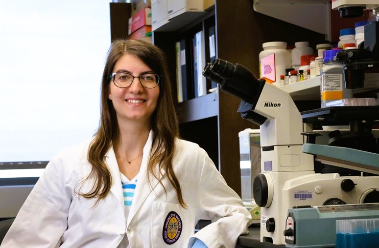
Giulia Monticone (Genetics)
Computational drug discovery for immunotherapy: Dr. Monticone is developing dual-functional small molecules that modulate the immune system to restore host defense. These compounds are designed to simultaneously target infectious agents or cancer cells and overcome immunosuppression, offering a powerful strategy for treating complex diseases such as viral infections and cancer. She has trained an AI-driven predictive model using experimental and literature-derived data to identify candidate molecules with dual immunomodulatory activity. She is leveraging this platform to design next-generation immunotherapeutics. This project combines computational drug discovery, immunology, and translational pharmacology to pioneer a new class of broad-spectrum, immune-restorative therapies. She collaborates with Dr. Dicle Yalcin.
Software utilized includes Seurat, Pymol and Alphafold.
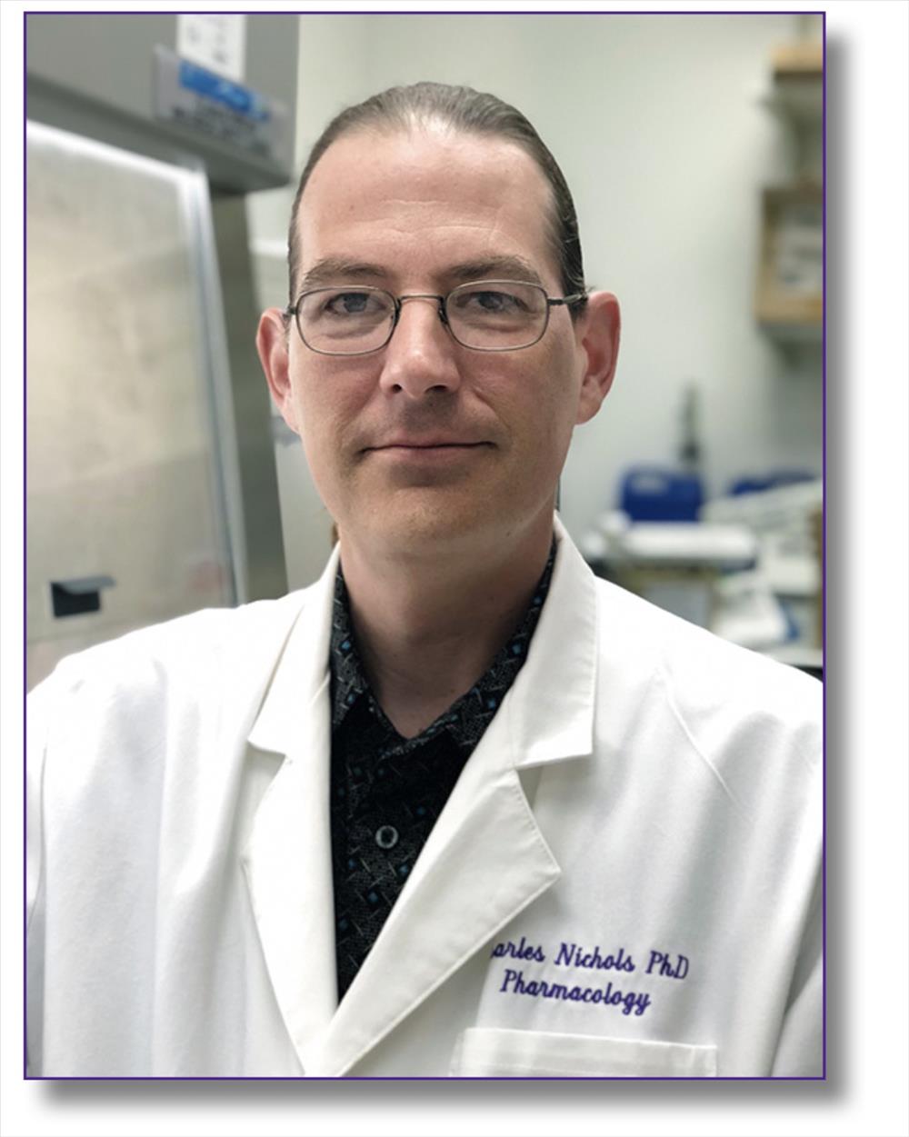
Charles Nichols (Pharmacology)
Dr. Nichols performs both genomic analysis (RNASeq and Single-Cell analysis(10x Cell Ranger)), and docking and molecular dynamics simulations of ligand/receptor interactions. Most of the time recently his workstation has been occupied by molecular dynamics simulations using Schrodinger Suite and Maestro/Desmond. He is currently running GROMACS molecular dynamics simulations on TigerFish.
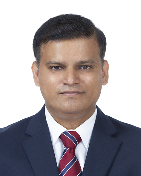
Anand Paul (Biostatistics and Data Science)
Dr. Paul develops resilient AI systems by integrating stochastic uncertainty modeling, and quantum statistical formulations to enable robust early-exit mechanisms. The framework achieves computational efficiency, adaptive robustness under noise, drift, and partial observability. Dr. Paul’s projects require scalable pipelines for multi-omics fusion, Bayesian/hierarchical modeling, and repeated cross-validation across cohorts.
One project, “The Area Deprivation Index and Revision Rates for Perioperative Joint Infection Following Primary Total Knee Arthroplasty” (Paul PI) is funded by the American Academy of Orthopedic Surgeons. This study investigates the relationship between neighborhood socioeconomic disadvantage—as quantified by the Area Deprivation Index (ADI)—and revision surgery rates due to perioperative joint infection (PJI) following primary total knee arthroplasty (TKA) in older adults. Utilizing data from the American Joint Replacement Registry (AJRR) and the 2022 ADI dataset, multivariable Cox proportional hazards models and logistic regression analyses will be applied to examine whether ADI independently predicts revision risk, while adjusting for clinical, demographic, and hospital-level covariates. By exploring both the direct effect of ADI and its mediating influence on known risk factors such as age, race, and sex, our study aims to elucidate the extent to which neighborhood-level disadvantage shapes postoperative outcomes.
Another NIH/NIAAA project is pending “Geroprotective Precision Medicine Strategies in PWH that Use Alcohol”. Dr. Paul is responsible for designing noise-tolerant ML pipelines that integrate clinical, behavioral, microbiome/metabolomic, and adherence data; emphasis on uncertainty quantification and outlier-resilient inference for individualized treatment selection. The project involves development of enhanced Hidden Markov Model sequence models and deep ensembles for longitudinal immune and biomarker trajectories, evaluation under domain shift and missingness, and targeted regularization and uplift modeling to estimate individual treatment effects (ITE) of probiotic/blueberry interventions on immune aging markers.
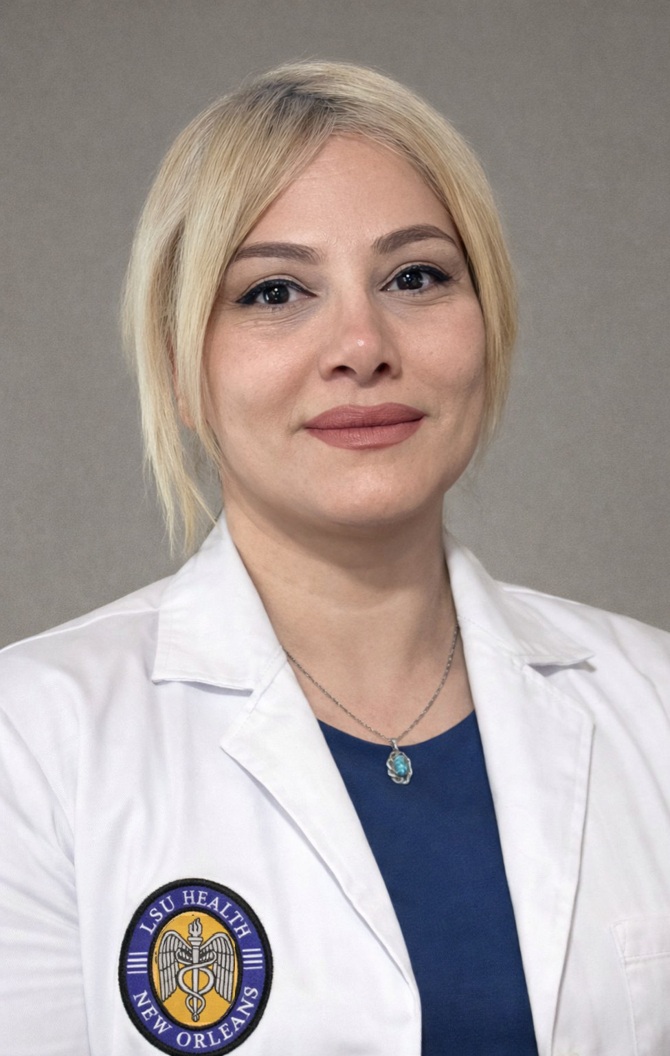
Vahideh Tarhriz (Cardiovascular Center)
Dr. Tarhiz was recently appointed faculty after receiving the 2026 American Heart Association (AHA) Career Development Award. As part of Center Director Dr. Lazartigues’ team, she has established a solid foundation in algorithms, machine learning, and bioinformatics. They are actively engaged in interdisciplinary research on gene expression and epigenetic modifications involving non-coding RNAs, in collaboration with computer scientists and software engineers. This collaboration aims to advance our capabilities in artificial intelligence, data analysis, and computational biology. They are currently working on several core areas of computer science, including: Data Structures and Algorithms, Operating Systems, Database Systems, Software Engineering. Dr. Tarhiz will implement a hybrid framework that combines Principal Component Analysis (PCA) for dimensionality reduction and a Multi-Layer Perceptron (MLP) for multi-label classification. This would enable experimentation with various models and evaluation of their performance using precision-based metrics such as F1-Score, Ranking Loss, and ROC-AUC.
Programming Languages: R language, Python, C++ , Databases: MySQL, PostgreSQL
Tools & Technologies: Git, Linux, Frameworks: TensorFlow, Pandas, NumPy
Business Intelligence Tools: Power BI
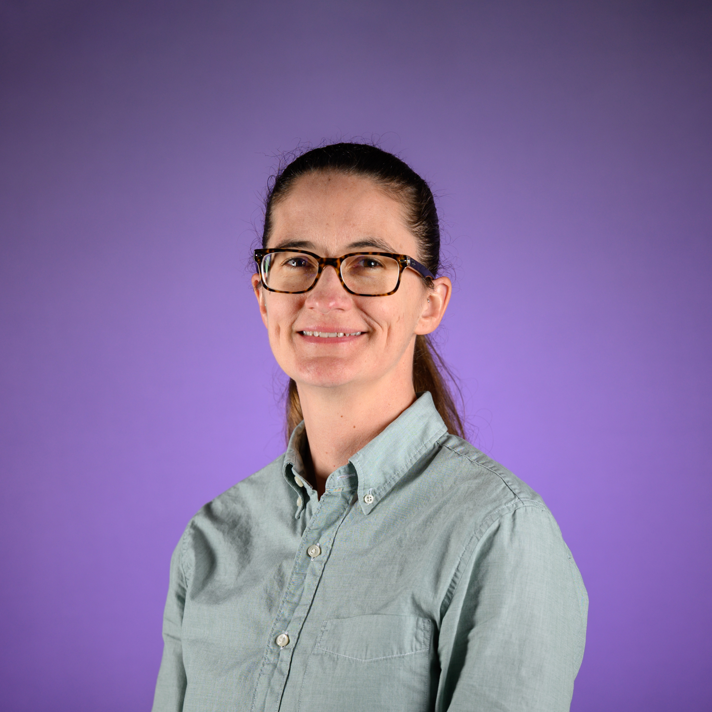
Carrie Wiese (Cardiovascular Center)
Dr. Wiese uses multiple -omic approaches to investigate gene regulatory mechanisms and the impact on cellular function and whole-body physiology related to cardiometabolic diseases, such as obesity, liver diseases, and atherosclerosis. Our multi-omics approach includes transcriptomics, lipidomics, proteomics, metabolomics, and epigenetic modifications (DNA methylation and histone modifications), with the goal of integrating these -omics datasets to understand the impact on disease development. This work requires high performance computing resources, including data storage infrastructure, secure data transfer, and parallelized computing for analysis, integration, and network generation for these datasets. This work is funded by American Heart Association Career Development Award (24CDA1269864), “Epigenetic Regulation in Atherosclerosis” 09/01/2014-08/31/2027.
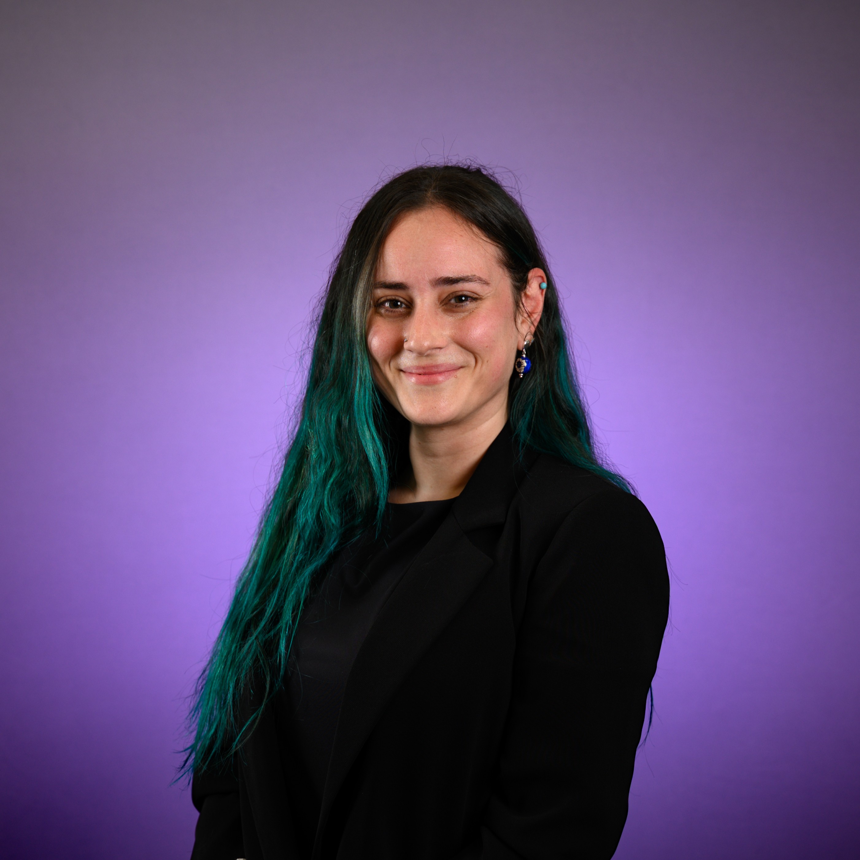
Dicle Yalcin (Interdisciplinary Oncology)
Dr. Yalcin’s work focuses on how prior and concurrent pathogen exposures shape antitumor immunity, with a particular emphasis on Kaposi sarcoma-associated herpesvirus (KSHV/HHV-8) and High-risk HPV-associated malignancies. To capture this complexity, she integrates multiple high-dimensional datasets spanning systemic and tissue compartments. They study 1) humoral responses against the human virome using high-throughput phage immunoprecipitation and sequencing in conjunction with Ag-specific B and T lymphocyte repertoire dynamics using high-avidity multimers and single-cell sequencing, 2) inflammatory and immunomodulatory pathways using proximity ligation assays (Olink), and 3) the tumor microenvironment using single-cell transcriptomics (Merscope) and proteomics (COMET) technologies. In parallel, Dr. Yalcin has working pipelines for building deep learning models for detection and quantification of Taupathy in brain specimens from a large cohort with AD pathology. Python, R, and MATLAB are used for most programming tasks. Analyses that require high-computing support and high-performance storage include:
- Antibody profiling and cross-reactivity: Handling millions of peptide Ag: Ab interactions, applying machine learning for epitope clustering, and integrating with demographic and clinical data.
- Single-cell multi-omics: Integration of TCR/BCR clonotypes and transcriptomic states using R packages such as Seurat, Signac, scRepertoire, immunarch, and CellChat, and Bayesian hierarchical modeling with brms and rstanarm for longitudinal outcome prediction.
- Large image files generated from Merscope and COMET spatial transcriptomics and proteomics. These datasets involve gigabyte- to terabyte-scale image stacks requiring GPU-accelerated rendering, scalable segmentation algorithms, and pipelines for integrating spatial context with molecular data. Tools include Fiji/ImageJ, QuPath, and Python frameworks (scanpy, squidpy, MONAI), alongside R-based spatial analysis packages such as Seurat, Giotto, and SpatialExperiment.
- Deep learning for image analysis: Convolutional neural networks and transformer-based architectures in Python (PyTorch, TensorFlow) enable segmentation, classification, and intensity quantification in brain IHC and tumor imaging datasets.
- HPC cluster access: To enable parallel processing of single-cell and repertoire data, antibody cross-reactivity computation from large alignments, and image quantification at scale.
- GPU resources: Essential for dimensionality reduction, spatial transcriptomics image embedding, deep learning–based segmentation, and integrative modeling across modalities. Large-memory nodes (≥512 GB RAM) for immune repertoire assembly and integration.
- Workflow generation: Reproducible pipelines (Nextflow, Snakemake) to connect multiple environments (R, Python, MATLAB, and proprietary pipelines like Cell Ranger/Space Ranger)